The key difference between fluorescence microscopy and confocal microscopy is that in fluorescence microscopy, the entire specimen is flooded evenly in light from a light source, whereas in confocal microscopy, only some points of the specimen are exposed to light from a light source.
Fluorescence microscopy is an important analytical technique that is useful in studying the properties of organic or inorganic substances. Confocal microscopy is an analytical technique useful in increasing the optical resolution and contrast of a micrograph using a spatial pinhole to block out-of-focus light in image formation.
CONTENTS
1. Overview and Key Difference
2. What is Fluorescence Microscopy
3. What is Confocal Microscopy
4. Fluorescence Microscopy vs Confocal Microscopy in Tabular Form
5. Summary – Fluorescence Microscopy vs Confocal Microscopy
What is Fluorescence Microscopy?
Fluorescence microscopy is an important analytical technique that is useful in studying the properties of organic or inorganic substances. The instrument that we use for this measurement is a fluorescence microscope. It is a type of optical microscope. This type of optical microscope uses fluorescence instead of scattering, reflection, attenuation, and absorption or in addition to them.
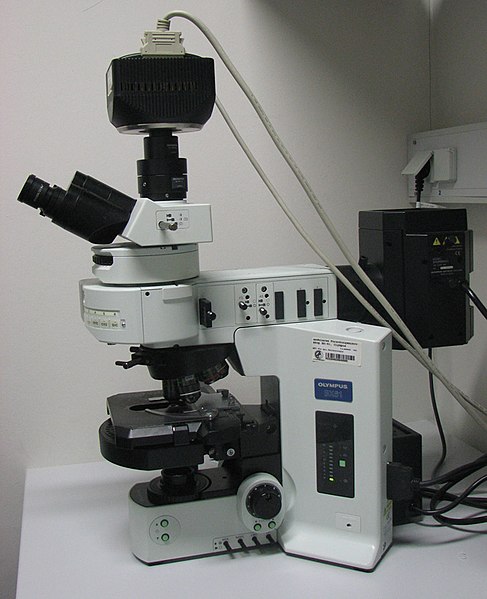
A fluorescence microscope uses fluorescence to generate an image. It might be a simple setup like an epifluorescence microscope or a more complicated design, including a confocal microscope that uses optical sectioning. This helps in getting a better resolution of the fluorescence image.

Figure 01: A Fluorescence Microscope
When considering the principle of fluorescence microscopy, we should use the specimen for illumination with light of a specific wavelength. There, the specimen absorbs light by fluorophores which causes them to emit light of longer wavelengths. It is important to separate the illuminated light from much weaker emitted fluorescence through the use of a spectral emission filter.
Typically, a fluorescence microscope contains a light source, excitation filter, dichroic mirror, and an emission filter. The light source can be a xenon arc lamp, mercury-vapor lamp, LEDs, and lasers. The dichroic mirror is also known as a dichroic beamsplitter.
What is Confocal Microscopy?
Confocal microscopy is an analytical technique useful in increasing the optical resolution and contrast of a micrograph using a spatial pinhole to block out-of-focus light in image formation. It is also known as confocal laser scanning microscopy or laser confocal scanning microscopy. It is an optical imaging technique.
In this microscopic technique, capturing multiple 2D images at different depths in a sample enables the reconstruction of 3D structures within an object. The confocal microscopic technique is useful extensively in scientific and industrial communities. It is typically used in life sciences, semiconductor inspection, and materials science.

Figure 02: The Confocal Point Sensor Principle from Minsky’s Patent
During the process, light travels through the sample under a conventional microscope as far into the specimen as it might penetrate, while a confocal microscope that only focuses a smaller beam of light passes through a narrow depth level at a time. This technique was developed by Marvin Minsky in 1957.
What is the Difference Between Fluorescence Microscopy and Confocal Microscopy?
Fluorescence microscopy is an important analytical technique that is useful in studying the properties of organic or inorganic substances. Confocal microscopy is an analytical technique useful in increasing the optical resolution and contrast of a micrograph using a spatial pinhole to block out-of-focus light in image formation. The key difference between fluorescence microscopy and confocal microscopy is that in fluorescence microscopy, the entire specimen is flooded evenly in light from a light source, whereas in confocal microscopy, only some points of the specimen are exposed to light from a light source.
The below infographic presents the differences between fluorescence microscopy and confocal microscopy in tabular form for side by side comparison.
Summary – Fluorescence Microscopy vs Confocal Microscopy
Fluorescence microscopy and confocal microscopy are two analytical techniques involved in optical imaging. The key difference between fluorescence microscopy and confocal microscopy is that in fluorescence microscopy, the entire specimen is flooded evenly in light from a light source, whereas in confocal microscopy, only some points of the specimen are exposed to light from a light source.
Reference:
1. “Fluorescence Microscopy.” An Overview | ScienceDirect Topics.
Image Courtesy:
1. “Olympus-BX61-fluorescence microscope” By Masur – Own work (CC BY-SA 3.0) via Commons Wikimedia
2. “Minsky Confocal Reflection Microscope” By Marvin Minsky (inventor), Ameter & Levy (Attorneys) – Clip of US Patent 3.013.467 (Public Domain) via Commons Wikimedia
ncG1vNJzZmivp6x7pbXFn5yrnZ6YsqOx07CcnqZemLyue9ahmK1lmah6tbTEZpuinpaav6a6wp5km52krLKmuoyfo66noprApLHNnJxmpZmYv7C%2FwqinsmWRo7Fur86nnaibkaF6rrXCq6asm5%2BlxnA%3D