Key Difference – Megakaryocyte vs Platelet
The process of blood clotting or thrombosis is mainly mediated by platelets in the blood. The blood clotting process is essential in order to prevent blood loss from the system during an external injury or an internal injury. Thus, it is important to maintain the count of platelets in the blood. It is essential to restore the platelet count immediately during conditions where platelet count is reduced. Megakaryocyte is the precursor of platelet cells, and it undergoes many intrinsic changes before being released as a platelet into the bloodstream. Platelets are a type of blood cell, required in the clotting process. This is the key difference between megakaryocyte and platelet.
CONTENTS
1. Overview and Key Difference
2. What is a Megakaryocyte
3. What is a Platelet
4. Similarities Between Megakaryocyte and Platelet
5. Side by Side Comparison – Megakaryocyte vs Platelet in Tabular Form
6. Summary
What is a Megakaryocyte?
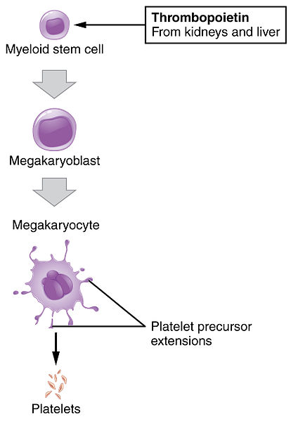
Megakaryocytes are nucleated, myeloid cells that are mainly found in the bone marrow, lungs and peripheral blood. Megakaryocytes have compact but lobed nuclei and a thin rim of basophilic cytoplasm and range up to 20 μm in size. The development of megakaryocytes occurs in a process called megakaryopoiesis which takes place in the fetal stage of an organism. Megakaryocytes arise from pluripotent hematopoietic stem cells and develop into two main precursor cells known as the burst forming cells and the colony forming cells. Then, megakaryocyte undergoes many intrinsic reactions to alter its cytoplasm and its membrane system in order to develop into a mature platelet. Out of the many factors aiding platelet formation from megakaryocytes, thrombopoietin (TPO) is the primary regulator involved. Upon complete maturation of the megakaryocyte, the cell is well equipped with all required proteins and other machinery required for platelet biogenesis.

Figure 01: Platelet Production from Megakaryocyte
Megakaryocytes produce extensions which participate in the process of releasing platelets to the blood. These protoplatelet extensions undergo another series of reactions to release the mature platelet to the blood.
What is a Platelet?
Platelet is a circulating anuclear disk shaped cells which account for nearly 20% of the total blood cell count. Its diameter lies between 3 to 4 μm. The mean normal platelet count is between 150,000 to 450,000 platelets per microliter of blood. The main function of platelets is to facilitate the process of blood clotting by forming platelet plugs in the first phase of the blood clotting process. Platelets also produce the platelet factor 3 which is important in the reaction process of coagulation. When the normal vascular integrity is disrupted due to an injury, circulating platelets and other factors assemble near the site of injury. Prostaglandins such as thromboxane aid the process of platelet aggregation and this is followed by the formation of the fibrin network on the site of injury to prevent further blood loss.
Disorders of platelets can cause many imbalances in the body. Certain health medications such as aspirin, which is a non steroidal anti-inflammatory drug, is administered to prevent blood clotting by interrupting a specific step of platelet aggregation.

Figure 02: Platelet
Genetic defects of platelet production are also currently under research in conditions like thrombocytopenia where reduced platelet number is quite common. Thrombocytopenia can be also resultant of certain viral infections such as Dengue, where the virus is capable of destructing platelets causing the platelet levels to decrease rapidly.
What are the Similarities Between Megakaryocyte and Platelet?
- The ultimate function of megakaryocytes and platelets is to initiate the blood clotting process.
- Organelle and glycoprotein subcellular distribution is similar in both cell types.
- The site of production of both cells is bone marrow.
What is the Difference Between Megakaryocyte and Platelet?
Megakaryocyte vs Platelet | |
| Megakaryocyte is a precursor of a platelet cell which is derived from a Hematopoietic stem cell. | Platelet is a type of blood cell involved in the clotting process. |
| Shape | |
| Size of a megakaryocyte is around 20 µm. | Size of a platelet is around 4-5 µm. |
| Structure | |
| Megakaryocytes are circular or ovoid shaped. | Platelets are disk-shaped flattened cells. |
| Presence of a Nucleus | |
| A nucleus is present in megakaryocyte. | A nucleus is absent in platelets. |
| Main Function | |
| The main function of megakaryocyte is the production of platelets and acting as platelet precursors | Blood clotting and initiating the coagulation process are the main functions of platelets. |
| Count per mm3 | |
| Megakaryocyte count is not specified. | Platelet count lies between 150,000 to 450,000 platelets per µl. |
Summary – Megakaryocyte vs Platelet
Blood clotting is a complex process which involves different types of cells. Megakaryocyte is the precursor of platelet cells, and it undergoes many intrinsic changes before being released as a platelet into the bloodstream. Platelets are a type of blood cell, required in the clotting process. This is the difference between megakaryocyte and platelet. The process of the transition from megakaryocyte stage to the mature platelet is a very complex process involving many factors. Even though the basic mechanisms of platelet maturation process have been elucidated, the specific regulatory mechanisms are yet to be understood and require further research.
Download PDF Version of Megakaryocyte vs Platelet
You can download PDF version of this article and use it for offline purposes as per citation note. Please download PDF version here Difference Between Megakaryocyte and Platelet
References:
1. Patel, Sunita R., et al. “The biogenesis of platelets from megakaryocyte proplatelets.” Journal of Clinical Investigation, American Society for Clinical Investigation, 1 Dec. 2005, Available here. Accessed 8 Sept. 2017.
2. Stiff, Patrick J. “Platelets.” Clinical Methods: The History, Physical, and Laboratory Examinations. 3rd edition., U.S. National Library of Medicine, 1 Jan. 1990, Available here. Accessed 8 Sept. 2017.
Image Courtesy:
1. “1908 Platelet Development” Connexions Web site. , Jun 19, 2013. (CC BY 3.0) via Commons Wikimedia
2. “Diagram of a platelet CRUK 407” By Cancer Research UK uploader – Own work (CC BY-SA 4.0) via Commons Wikimedia
ncG1vNJzZmivp6x7pbXFn5yrnZ6YsqOx07CcnqZemLyue8OinZ%2Bdopq7pLGMm5ytr5Wau265xKCYpJmirrykxdOeZJqmlGLDtHnPpZitnZyawXA%3D